Key Points for Culturing B16 Cell Line
Source: PricellaPublished: 2024-05-08
B16 cell line is a murine melanoma cell line derived from C57 mice, commonly used in tumor biology research.
In this article, we have compiled the methods and precautions for culturing B16 cell line, as well as the causes and solutions for melanin formation in B16 cells, to help you quickly get started with your experiments.
1.Culturing Methods for B16 Cells
-
Culture Conditions
RPMI-1640 medium (Cat.No.: PM150110) supplemented with 10% fetal bovine serum (FBS) and 1% penicillin-streptomycin (P/S) (Cat.No.: PB180120).
Or B16 Cells Complete Medium (Cat.No.:CM-0029)
-
Growth Characteristics
Adherent growth with rapid proliferation and the ability to produce melanin.
-
Passaging Method
Dissociate the cells with 0.25% trypsin-EDTA for 2-3 minutes. It is recommended to passage the cells at a ratio of 1:3 to 1:4.
-
Cryopreservation
Recommended freezing density is 1-5 × 10^6 cells/mL.
Precautions
1)Cell Density: As the cells grow rapidly, maintain a moderate culture density. Regular passaging and medium changes are essential. Overly high cell density can result in poor cell - condition and potential cell death.
2)Melanin Content: The cells contain melanin, which may cause the culture medium to darken in color. When dissociating and centrifuging the cells, the cell pellet may appear black-brown or gray-black, which is a normal phenomenon.

Fig.1 When reviving the cells, the cell pellet may appear gray-black in color when using RPMI-1640 culture medium.

Fig.2 The production of melanin by the cells can lead to a change in the color of the culture medium, such as in RPMI-1640 medium.
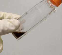
Fig.3 In DMEM culture medium, the production of melanin by the cells causes a change in the color.
2.Causes of Melanin Formation
The B16 cell line can be cultured in vitro under two distinct conditions: rapid proliferation or differentiation and maturation. Varying the culture medium can change the growth state of the cells. RPMI-1640 medium promotes rapid proliferation, while DMEM medium encourages cell differentiation and maturation, which leads to melanin synthesis. For routine expansion and cryopreservation, RPMI-1640 medium is recommended. To conduct experiments involving melanin secretion or other related studies, switch to DMEM medium after expanding and cryopreserving the cells to meet the experimental design.
It is important to note that B16 cells are sensitive to culture medium conditions. Even when using the same DMEM or RPMI-1640 medium, slight differences in specific components from different models or manufacturers can change the growth state of the B16 cells. Careful selection of culture medium is advised during experiments, and it is best to use the same model of culture medium from the cell supplier to ensure stable cell characteristics.
Changing culture conditions, such as switching from RPMI-1640 to DMEM, can lead to significant melanin formation and blackening of the culture medium. However, if you observe the culture medium suddenly turning black without switching culture mediums during stable culture, the possible causes could include:
(1) Cell Death
If there is significant cell death during the culturing process, metabolic byproducts and cell debris can cause the culture medium to turn black. This may occur due to poor cell condition, contamination, or other factors.
Solution: Monitor the condition of the cells and ensure that the growth conditions and culture medium are appropriate. If excessive cell death is observed, you may need to revive the cells from a cryopreserved stock.
(2) Cell Contamination
If contamination occurs in the culture medium (bacteria, fungi, mycoplasma, or nanobacteria), the growth and metabolic byproducts of these microorganisms can lead to the darkening of the culture medium.
Solution: Investigate the source of contamination and inspect the culturing environment, including the reagents and consumables used. Practice strict aseptic techniques. If contamination is detected, immediately replace the culture medium, perform sterile cleaning, and eliminate the source of contamination.
(3)Customer Experience with B16-F10 Cells
(The following is a shared experience from our customers and is only for reference.)
-
Trypsinization:
To maintain the viability of B16-F10 cells, it is advisable to carefully control the temperature and duration of trypsinization during passaging. Monitor the dissociation state of the cells (under a microscope, you should observe cells detaching and rounding up, and the cells should slide off the side of the culture flask when tilted, indicating that dissociation can be terminated). Over-dissociation can result in cell damage and decreased viability, especially when performing experiments related to melanin synthesis, secretion, and tyrosinase activity. Pay close attention to the trypsinization time and the state of the cells.
-
Irradiation with Light:
There are two key points to pay attention to when irradiating B16-F10 cells with light:
I. Ensure that the culture dish lid is open during irradiation so that the light reaches the B16-F10 cells directly, rather than being refracted or absorbed by the dish lid.
II. Before irradiation, replace the medium with RPMI-1640 complete medium without phenol red. This prevents the phenol red from absorbing light and interfering with experimental results.
(4)Common Applications of B16 Cell Line
A.Melanoma Biology Research
B16 cell line can be used to study the mechanisms of melanoma initiation, progression, and metastasis. By simulating the behavior of melanoma cells in vivo, researchers can explore processes such as cell proliferation, migration, invasion, and metastasis.
B.Tumor Drug Screening
B16 cells cultured in vitro can be used to screen and evaluate the efficacy of anti-melanoma drugs. By adding drugs to the culture medium, researchers can study the effects of the drugs on cell proliferation, apoptosis, invasion, and migration to assess their antitumor activity.
C.Immunotherapy Research
B16 cells can serve as a model for immunotherapy research. For example, researchers can add immune stimulants or immune checkpoint inhibitors to the culture medium to study their effects on the immune response and tumor growth, assessing their potential in melanoma treatment.
D.Melanoma Metastasis Research
The metastatic capability of B16 cells is an important aspect of their research applications. By injecting the cells into the bloodstream or specific organs of mice, researchers can study the mechanisms of melanoma cell migration and metastasis, as well as therapeutic strategies targeting metastasis.
E.Cell Signaling Pathway Research
B16 cells can be used to investigate the role of cell signaling pathways in the initiation and progression of melanoma. For example, by transfecting or inhibiting specific signaling molecules or pathways, researchers can study their effects on biological behaviors such as cell proliferation, apoptosis, and invasion.
Note:
While B16 cell line is widely used in melanoma research, there are limitations to its use in in vitro culture, such as differences from the in vivo environment and the heterogeneity of the cell line. Therefore, researchers should use a combination of other models and methods to better simulate and understand the biological characteristics of melanoma.
References:
[1]Bae, J. S., Han, M., Yao, C.,et al. Chaetocin inhibits IBMX-induced melanogenesis in B16F10 mouse melanoma cells through activation of ERK. Chemico-biological interactions,2016,245, 66–71. https://doi.org/10.1016/j.cbi.2015.12.021.
[3]Costantino, V. V., Lobos-Gonzalez, L., Ibañez, J.,et al.Dehydroleucodine inhibits tumor growth in a preclinical melanoma model by inducing cell cycle arrest, senescence and apoptosis. Cancer letters,2016,372(1),10–23.https://doi.org/10.1016/j.canlet.2015.12.004.
[4]Chen, J. H., Chang, J. L., Chen, P. R.,et al. Inhibition of peroxisome proliferator-activated receptor gamma prevents the melanogenesis in murine B16/F10 melanoma cells. BioMed research international, 2014, 695797. https://doi.org/10.1155/2014/695797.
Prev: A Comprehensive Overview of A-498 Cells
Next: How to Successfully Culture SH-SY5Y Cells: Key Details to Consider


