Unraveling the Mystery of Cytoplasmic Vacuoles
Source: PricellaPublished: 2025-02-18
Vacuoles in cells are bubble-like structures that are easily visible under a light microscope due to their optical properties, which contrast sharply with the surrounding cytoplasm. When such distinctive vacuoles appear within cells, the phenomenon is referred to as cytoplasmic vacuolization.
In this article, we will explore the causes of cytoplasmic vacuolization and discuss practical strategies to address the issue.
What Are Vacuoles and Where Do They Come From?
Cytoplasmic vacuoles originate from a variety of intracellular membrane-bound organelles, including mitochondria, the endoplasmic reticulum (ER), lysosomes, and the Golgi apparatus. Under specific physiological or pathological conditions, these organelles may undergo complex biochemical processes, resulting in the formation of vacuoles within the cell.

Figure 1. Cytoplasmic vacuoles under a light microscope
Causes of Cytoplasmic Vacuolization and How to Address Them
1.Cellular Senescence
Cytoplasmic vacuolization is commonly observed during the senescence of primary cells in culture. Primary cells have a limited capacity for proliferation in vitro. When cultured for extended periods, these cells undergo spontaneous aging, leading to vacuole formation and, in some cases, cell death. Similarly, continuous cell lines may also develop vacuoles as a result of cellular senescence due to excessive passaging.
Solution:
For primary cells, use cells at lower passage numbers and avoid prolonged in vitro culture to
• reduce the risk of aging and vacuole formation.
• For cell lines, strictly control the number of passages and avoid using high-passage cells to prevent senescence-related vacuolization.
2.Changes in Media, Serum, or Culture Conditions
Switching culture media or serum can stress cells, leading to vacuole formation. Additionally, improperly prepared or stored media, expired reagents, or low-quality serum can alter the physicochemical properties of the culture environment (e.g., pH, osmolarity), negatively impacting cell health and potentially causing vacuolization.
Solution:
When receiving new cells, avoid an abrupt switch to new media or serum. Gradually transition by mixing the original media with the new media to minimize stress on the cells.
Always prepare fresh media, adjust pH and osmolarity to optimal levels, and ensure the use of high-quality serum. Consider using Pricella® cell culture media, which offers a wide range of rigorously quality-controlled products and customizable options to mitigate vacuolization risks caused by media-related issues.
3.Improper Cell Dissociation
For adherent cells, improper dissociation during passaging—such as using the wrong dissociation reagent, over-digesting, or applying excessive mechanical force—can damage cells and lead to vacuole formation.
Solution:
Select appropriate dissociation reagents tailored to your cell type. Carefully monitor dissociation time and handle cells gently to minimize damage. Pricella® provides a wide selection of dissociation reagents to meet your specific cell culture needs.
4.Delayed Passaging or Media Changes
Failure to passage confluent cells or perform timely media changes can result in the accumulation of metabolic byproducts and depletion of nutrients in the culture environment. These conditions may induce autophagy, leading to vacuole formation.
Solution:
• Regularly monitor cell growth and passage cells when they reach approximately 80% confluency.
• Replace the culture media every 2-3 days with fresh media to maintain an optimal environment.
5.Contamination
Contamination with bacteria, mycoplasma, or nanobacteria can severely compromise cell health, leading to vacuolization and other abnormal phenomena.
Solution:
Follow strict aseptic techniques during cell culture. Use sterile reagents and consumables to minimize contamination risks. For less obvious contaminants like mycoplasma, perform routine screening and consider conducting empty culture (media-only) tests to detect hidden issues. Address contamination immediately to prevent it from spreading.
6.Drug-Induced Effects
During experiments, researchers often treat cells with specific drugs (e.g., chloroquine, aminoglycoside antibiotics, apoptosis inducers). While these drugs may induce desired biological effects, they can also be cytotoxic, potentially causing vacuole formation or even cell death.
Solution:
Carefully optimize drug concentrations and treatment durations according to experimental requirements to minimize cytotoxicity.
Can Cytoplasmic Vacuolization Be Reversed?
The reversibility of vacuole formation largely depends on its underlying cause and the cell’s overall condition:
• Reversible Vacuolization: If vacuoles are induced by short-term environmental changes (e.g., temperature fluctuations or transport-related stress), cells may recover when unfavorable conditions are removed, and the culture environment is restored.
• For example, cells shipped at ambient temperature may develop vacuoles due to transport stress (Figure 2). However, these vacuoles often disappear after cells are stabilized and subcultured under normal conditions (Figure 3).
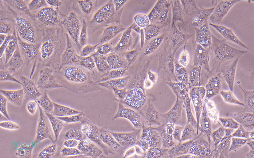
Figure 2. Vacuoles in MC3T3-E1 Subclone 14 cells upon arrival after transport stress
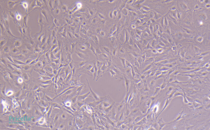
Figure 3. MC3T3-E1 Subclone 14 cells after stabilization and passaging
• Irreversible Vacuolization: When vacuoles form due to severe cellular damage—such as prolonged culture under suboptimal conditions, contamination, or significant structural and functional impairment—they are often irreversible. In these cases, the vacuolization process usually leads to cell death.
Prev: A Comprehensive Guide to Detecting and Handling Laboratory Contamination
Next: Five Common Causes of Cell Elongation and How to Address Them


