Understanding Abnormal Morphology of Renca Cells
Source: PricellaPublished: 2024-10-23
Introduction
Renca (mouse renal carcinoma) cells are epithelial cells isolated from the kidneys of male mice with renal cortical adenocarcinoma. The growth pattern of these cells closely resembles that of adult renal carcinoma, particularly in their spontaneous metastasis to the lungs and liver. Notably, Renca cells do not express the TGF-b type II receptor, making them a common choice for establishing Renca tumor models in nude mice.
Issue: Abnormal Cell Morphology
A researcher recently observed a discrepancy in the morphology of their Renca cells compared to the standard morphology documented by authoritative cell banks. This raised concerns about the authenticity of the cells in question. If the cells you are culturing resemble those in Figure 1, and appear significantly different from the cells shown by the cell bank, yet STR (short tandem repeat) profiling confirms them as Renca cells, what could be the cause of this morphological difference? The likely culprit may be the serum used during culture.
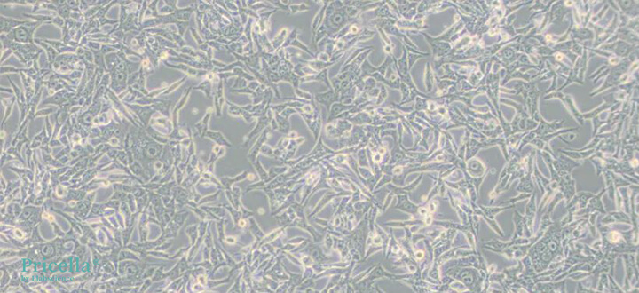
Figure 1. Renca cells cultured for 2-3 days after passage
The Impact of Serum on Cell Morphology
Renca cells are notably sensitive to serum quality. Changes in serum composition can lead to alterations in cell morphology. If the cells exhibit normal growth but undergo morphological changes after a serum change, it is possible that the cells are still adjusting to the new conditions. Given time to adapt, the cells may return to their typical morphology.
Therefore, when encountering abnormal morphology in Renca cells, it is advisable to remain calm and continue passaging the cells. The cells may recover after several passages.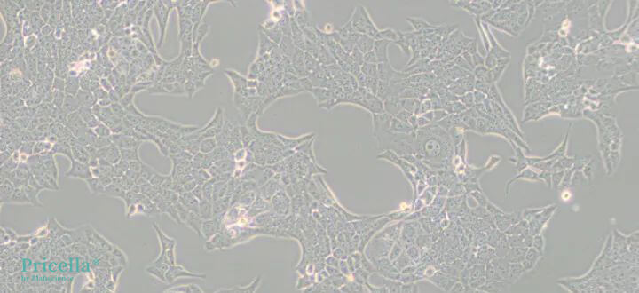
Figure 2. Renca cells with adjusted morphology
Recommendation
When altering cell culture conditions, it is prudent to first test a small sample of cells to ensure compatibility.
Important Considerations
Renca cells are sensitive to serum quality, which significantly influences their morphology. Always select high-quality serum for culturing these cells.
The growth medium for Renca cells should be RPMI-1640 supplemented with 10% FBS, 1% P/S, 0.1mM NEAA, 1mM Sodium Pyruvate, and 2mM L-glutamine.
Variations in medium and serum batches can also lead to morphological changes in Renca cells. Minimize batch changes whenever possible, and conduct small-scale tests before implementing any changes.Prev: Technical Principles and Procedures for Cell Cryopreservation and Resuscitation
Next: CELL BIOLOGY ACADEMY | ULTRA-DETAILED APOPTOSIS ASSAY SELECTION GUIDE!


