Troubleshooting Precipitation in Cell Culture: Causes and Solutions
Source: PricellaPublished: 2024-04-09
Cell culture is an essential skill for every researcher. As the saying goes, 'Good cell growth, no experimental troubles.' However, the reality often shows that the cells in the laboratory are prone to 'illness.' While others' culture mediums are pink and clear, with cells thriving, we often encounter various turbidity and precipitation issues for reasons unknown during cell culture. What could be causing this situation?
There are two main reasons for the occurrence of precipitation during cell culture:
- Cell contamination itself;
- Precipitation of metals, proteins, and other components of the culture medium within the medium.
First, let's take a look at cell contamination.
1.Bacterial/Fungal/Yeast Contamination
Bacterial, fungal, and yeast contaminations are relatively easy to detect because they can be identified by the naked eye and under a microscope, as shown in the figure below:
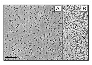
Figure 1. Bacterial Contamination
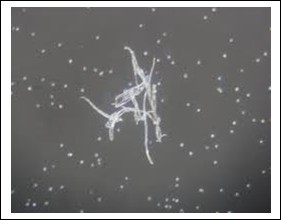
Figure 2. Fungal Contamination
Among these three types of contamination, bacterial contamination stands out for its distinct characteristics and can be somewhat challenging to identify. Common bacteria include rod-shaped bacteria, cocci, and spiral-shaped bacteria.
2.Mycoplasma Contamination
Mycoplasma is the nemesis of cells because its average diameter is only 0.1 to 0.3 µm, making it undetectable under a microscope.
Furthermore, mycoplasma contamination exhibits slow initial growth, which cannot be observed with the naked eye.
Detection: The commonly used detection method is the culture method, where 1 mL of cell culture medium is added to a liquid medium containing mycoplasma and cultured for 5 days. The color change of the medium is observed during this time. If it turns yellow, it is suspected that the cells are contaminated with mycoplasma. If there is no color change, then blindly passaged for one generation.
If the medium turns yellow, mycoplasma solid culture medium can be used. It is cultured in a 5% CO2 incubator for 3 to 5 days, and the presence of "fried egg"-shaped mycoplasma colonies is observed. Once contamination occurs, both the cells and the culture medium should be discarded.
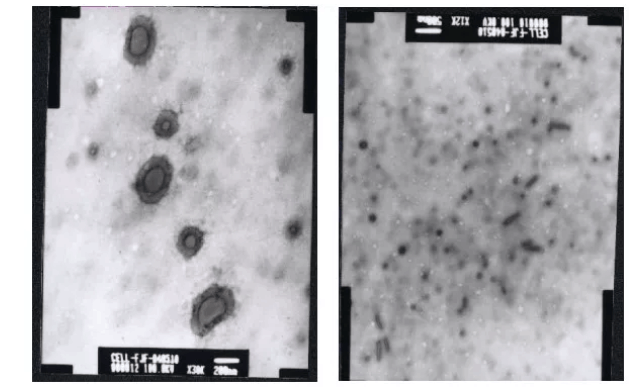
Figure 3. Mycoplasma Contamination (Left: Electron Microscope Image (×30k); Right: Electron Microscope Image (×12k))
In addition, mycoplasma contamination can also be detected using methods such as fluorescent staining and phase-contrast microscopy observation, or by directly purchasing mycoplasma detection kits for testing.
Treatment Measures:
- Regularly perform mycoplasma testing using mycoplasma detection kits.
- Changing the medium can slow down contamination but cannot eliminate it.
- Anti-Mycoplasma treatment kits can achieve better results.
- Macrophage clearance from the peritoneal cavity of mice.
Recommendation: Pricella Mycoplasma Removal Medium (Catalog Number: PM150110-MR)
Pricella Mycoplasma Removal Medium contains special ingredients designed to eliminate mycoplasma contamination. This unique formulation, a combination of compounds, achieves effective mycoplasma elimination by inhibiting the synthesis of DNA and proteins necessary for mycoplasma growth. It aims to salvage valuable cells and minimize losses caused by mycoplasma contamination.
This product has undergone testing on numerous cell types and extensive experimental validation. It is proven to be non-toxic to cells and demonstrates significant efficacy in eliminating mycoplasma contamination. It effectively inhibits and kills mycoplasma, as illustrated below:

Figure 4. Cells after treatment with Pricella Mycoplasma Removal Medium
3. Contamination by Eukaryotic Cells
Eukaryotic cell contamination refers to the presence of other cells mixed into a cell line. Due to the subtle differences in the size and shape of various cells, this type of contamination is also difficult to identify.
In general, when faced with this situation, the only solution is to discard the contaminated cells because this contamination can significantly affect the reproducibility of experimental results.
Next, let's address the issue with the culture medium.
If contamination is ruled out, the turbidity in the cell culture medium is typically attributed to the precipitation of metals, proteins, and other components of the culture medium. These precipitates may compromise cell health as they can chelate and remove essential nutrients and other necessary components, thereby altering the composition of the medium.
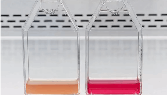
Figure 5.
Possible causes of precipitation and solutions:
1.Temperature Fluctuations
In cell culture, temperature is one of the main factors causing precipitation. When exposed to extreme temperature changes, high molecular weight plasma proteins may precipitate from the solution.
Solution: It is essential to carefully follow the recommended guidelines for selecting the optimal storage and handling methods for cell culture media. Additionally, avoid repeated freeze-thaw cycles to prevent precipitation.
2. Concentration Changes Induced by Dehydration
If the culture medium is prone to evaporation, the concentration of medium components (including salts) will increase, especially at the surface of the medium where crystal precipitates may form.
Solution:
- Ensure that culture bottles/plates do not undergo evaporation by monitoring the humidity within the incubator.
- Seal culture vessels to prevent dehydration.
3. Calcium Salts
When preparing serum-free culture media, the order of component addition may lead to the formation of insoluble molecules. Calcium salts are particularly prone to precipitation; for instance, CaCl2 and MgSO4 can react in solution to produce CaSO4 crystals. High-pressure sterilization and pH instability can exacerbate this issue.
- Dissolve CaCl2 separately in deionized water before adding other culture medium components.
- Adding buffering agents can help maintain osmotic pressure stability.
4. Metal Supplements
Metal ions such as copper, iron, and zinc are essential for cell growth and are crucial supplements in serum-free culture media.
The absence of other serum components in the culture system may lead to the precipitation of these metals, creating a toxic environment for cells.
Under higher pH conditions (generally >8), carbonate and hydroxide ions can form insoluble precipitates with copper ions, phosphate and hydroxide ions with iron ions, carbonate, phosphate, and hydroxide ions with zinc ions, and hydroxide ions with magnesium ions.
Under oxidative conditions, copper and zinc are more prone to precipitation.
Metal Precipitate Color Chart:
| Color | Precipitate | Color | Precipitate |
| Black | Copper(I) sulfide | Red-brown | Iron(III) hydroxide |
| Black | Copper(II) oxide | Rose | Manganese carbonate |
| Black | Iron(II) oxide | Steel-gray | Zinc phosphate |
| Black | Iron(II) sulfide | White | Calcium phosphate |
| Blue | Copper(II) hydroxide | White | Magnesium carbonate |
| Blue | Iron(II) phosphate | White | Magnesium hydroxide |
| Blue-gray | Copper(II) cysteinate | White | Magnesium peroxide |
| Blue-dark green | Copper(II) carbonate | White | Magnesium pyrophosphate |
| Blue-green | Copper(II) chloride oxide | White | Zinc carbonate |
| Brown | Iron(III) acetate | White | Zinc oxide |
| Colorless | Calcium carbonate | White | Zinc sulfide |
| Colorless | Zinc hydroxide | Yellow | Copper(I) carbonate |
| Gray-black | Copper(I) phosphide | Yellow | Copper(I) hydroxide |
| Gray-green or brown-black | Magnesium oxide | Yellow | Zinc peroxide |
| Red-black | Iron(III) oxide |
Prev: Common Types of Cell Culture Contamination
Next: How to Rescue Cells with Slow Growth and Poor Adhesion in Passages?


