Common Types of Cell Culture Contamination
Source: PricellaPublished: 2024-04-08
Components detrimental to cell survival and resulting in impurities within the cell culture environment should be classified as cell contamination. While complete elimination of cell contamination is unattainable, its frequency and severity of impact can be mitigated.
This article will briefly introduce several common types of cell contamination and their handling methods.
Common Types of Cell Culture Contamination
-
Bacterial Contamination
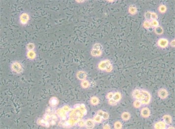
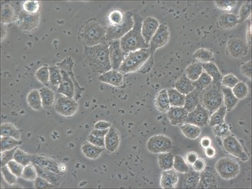
Characteristics: Bacteria typically range in size from 0.5 to 5 μm, much smaller than animal cells. They are often spherical or rod-shaped, with regular morphology and uniform distribution. Bacteria proliferate rapidly, with an explosive increase typically occurring 48 to 72 hours after contamination. They consume nutrients quickly, leading to rapid yellowing and cloudiness of the culture medium. Under the microscope, bacteria exhibit obvious directional movement.
Treatment: Mild bacterial contamination can be addressed with a 10X penicillin-streptomycin solution (Cat. No.: PB180120) washing treatment. For severe bacterial contamination, it is advisable to discard the contaminated materials after disinfection.

(Figure 1)
(Figure 2)
-
Fungal Contamination
Characteristics: Fungal contamination manifests as chain-like or filamentous structures, often forming visible colonies. It may not induce any color change or may result in a purple hue in the culture medium. This type of contamination predominantly impacts local cells, while other areas continue to grow unaffected. Fungi have the capability to produce spores, facilitating easy contamination of neighboring cells.
Treatment: Eradicating fungal contamination proves challenging and retaining contaminated materials is not advisable. After disinfection, it is recommended to discard the contaminated materials.

(Figure 3)

(Figure 4)
-
Mycoplasma Contamination
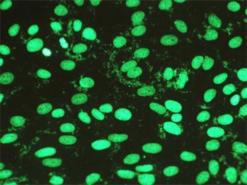
Characteristics: Mycoplasma is the smallest microorganism, invisible under an optical microscope but observable through an electron microscope. Cells infected with mycoplasma exhibit a sudden increase in debris at a certain time point, and with successive passages, the cellular condition significantly deteriorates. Mycoplasma contamination can spread through aerosols, affecting other cells in the same cell culture room.
Identification methods: PCR (Polymerase Chain Reaction), staining methods, fluorescence staining, etc.
Treatment: If the cells remain viable after contamination, culturing them with anti-Mycoplasma treatment reagent (Cat. No.: P-CMR-001) for 2-3 passages can lead to mycoplasma clearance. If the cells exhibit severe deterioration after contamination, it is advisable to discard them after disinfection.
(Figure 5)
(Figure 6)

(Figure 7)

(Figure 8)
-
Nanobacteria Contamination
Characteristics: Nanobacteria contamination lacks a distinct definition and cannot be directly observed under a microscope. It is primarily characterized by poor cell condition, presence of black spots and debris, and Brownian motion. Detection reveals mainly mycoplasma contamination with some instances of proliferation by unidentified microorganisms.
Treatment: If the cells remain viable after contamination, culturing them with anti-nanobacteria treatment reagent (Cat. No.: P-CMR-002)/ anti-Mycoplasma treatment reagent (Cat. No.: P-CMR-001) for 2-3 passages can lead to clearance. If the cells exhibit severe deterioration after contamination, it is advisable to discard them after disinfection.
-
Cross-Contamination
Characteristics: Cross-contamination refers to the mixing of different cell types together.
Identification Methods:
For the same species: Short Tandem Repeat (STR) analysis, with more than 3 alleles. (Consideration of the cell's genetic background is necessary, such as EA.hy926 cells, which are hybrid cells containing genetic information from two sources.)
For different species: PCR (Polymerase Chain Reaction) method, amplifying species-specific genes where products of different species have different sizes.
Treatment: It is recommended to reseed the culture with authentic cells.

(Figure 9)
Dealing with Cell Contamination
Once cell contamination is detected, it's essential to promptly clean all reagents and consumables used in the cell culture process. Consumables can be replaced with new ones or subjected to high-pressure sterilization. Reagents can be refiltered for sterility or replaced with a new batch. Workbenches and cell culture rooms should undergo comprehensive cleaning and disinfection with appropriate disinfectants before introducing new cells to prevent recurring contamination incidents.
Strategies for Preventing and Managing Cell Contamination
NO.1
Cell Culture Room Environmental Control
Environmental Requirements:
Clean area: Class 10,000
Local area: Class 100
Regular monitoring for settling bacteria
Environmental Disinfection: Weekly scheduled disinfection and sterilization
1)Floors/Walls: Use disinfectants to clean the floors and walls
2)Environmental Disinfection: Regular UV irradiation, ozone fumigation
3)Instrument and equipment surfaces: Wipe with 75% alcohol
NO.2
Management of Laboratory Personnel
Attire:Wear coveralls, masks, and gloves, ensuring hair and wrists are covered to minimize exposed skin.
Behavior: Minimize movement and door opening/closing, avoid talking during operations.
Skills: Develop good aseptic technique habits.
Others: Reduce shared items, establish uniform practices in common areas, differentiate between clean and high-risk areas.
NO.3
Management of Laboratory Supplies
Reagents: Ensure sterility before use; self-prepared reagents should be sterilized by filtration before use.
Consumables: For reusable consumables, adhere to prescribed cleaning and autoclaving procedures, using sterilization indicators. Once sterilized and dried, place consumables immediately in a laminar flow hood. Use within one week; any unused items must be re-sterilized before reuse.
Instruments and Equipment: Regularly wipe and disinfect instruments; high-risk areas like incubators and microscope trays require frequent cleaning. Add antimicrobial agents to water trays in incubators and water baths used for cell recovery.
Others: Promptly clean up spills in cell cultures; in case of localized contamination, identify the source of cell contamination and conduct overall environmental disinfection.
Prev: Decoding Cell Digestion:Are Your Cells Ready?
Next: Troubleshooting Precipitation in Cell Culture: Causes and Solutions


