Comprehensive Overview of Astrocyte Knowledge - Pricella
Source: PricellaPublished: 2025-01-02
Astrocytes (AST) are the largest type of glial cells in the mammalian brain, constituting 20-40% of all glial cells. These cells play crucial roles in the central nervous system (CNS), including synapse formation, maintaining ion homeostasis, neurotransmitter regulation, and the formation and maintenance of the blood-brain barrier (BBB).
In this session, we will provide a summary of astrocyte classification, identification, primary isolation, and culture methods, along with key precautions to help you get started quickly with astrocyte-related experiments.
Classification of Astrocytes
Traditionally, astrocytes are classified into two types:
1. Fibrous Astrocytes
Primarily distributed in white matter. These cells have elongated processes with fewer branches and contain a high amount of glial filaments in the cytoplasm. The main component of these glial filaments is glial fibrillary acidic protein (GFAP).
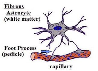
Fibrous Astrocytes(The image is sourced from the internet)
2. Protoplasmic Astrocytes
Mainly distributed in grey matter. These cells have shorter, thicker processes with more branching and less glial filament content in the cytoplasm.
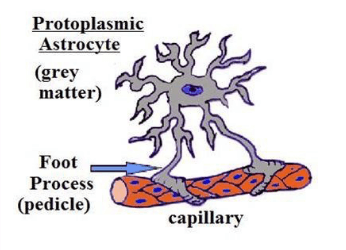
Protoplasmic Astrocytes(The image is sourced from the internet)
3. Morphology and Identification
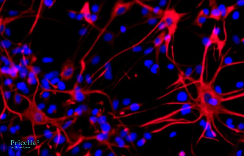
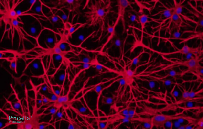
Rat Astrocyte Identification Image Provided by Pricella®
GFAP is a cytoskeletal protein specific to astrocytes. During early embryonic neural development, astrocyte precursors mainly express vimentin. As astrocytes mature, vimentin expression decreases while GFAP expression increases, continuing throughout the cell's lifespan. As such, GFAP is widely recognized as a hallmark marker of astrocytes. Under normal physiological conditions, GFAP expression is low in the brain, but it becomes upregulated in response to pathological conditions, though the exact mechanisms remain unclear.
Comparison Between Rodent and Human Protoplasmic Astrocytes
A: Typical mouse protoplasmic astrocyte. GFAP (white). Scale bar = 20 μm.
B: Typical human protoplasmic astrocyte at the same scale. Scale bar = 20 μm.
C and D: Human protoplasmic astrocytes are 2.55 times larger than mouse astrocytes.
E: Mouse protoplasmic astrocytes stained with DiI (white) and Sytox (blue), displaying the full structure of astrocytes, including their numerous fine processes. Scale bar = 20 μm.
F: 3D visualization of human astrocytes reveals a highly intricate network of fine processes that define the complexity of human protoplasmic astrocytes. Scale bar = 20 μm.
4. Primary Isolation
1) Isolate cells from brain tissue of 1-3 day-old neonatal mice.
2) Dissociate brain cells using 0.25% trypsin.
3) After obtaining a mixed cell population, centrifuge at 300×g for 10 minutes multiple times to reduce contamination from non-astrocytic cells.
4) Perform differential adhesion for 30-60 minutes to remove fibroblasts.
5) During subsequent culture, shake the cells at each medium change to further purify the astrocyte population.
Culture Considerations
Astrocytes can be cultured using standard adherent cell culture methods. Below are three key considerations for culturing astrocytes:
1) Standard 0.25% trypsin can be used for dissociation.
2) Passage: Cells can typically be passaged up to 3 generations, although cell morphology may change slightly with prolonged passaging.
3) Nutrient Requirements: Astrocytes have low nutrient demands and can grow in normal complete growth media. Adding nerve growth factor (NGF) can promote better maintenance of cell morphology and proliferation.
Prev: Essential Techniques for Accurate Cell Counting in the Lab - Pricella


