A Comprehensive Guide to Epithelial Cell Research: Structure, Function, Isolation & Culture Techniques
Source: PricellaPublished: 2025-05-14
Epithelial cells form continuous layers that cover the body's external surfaces and line internal cavities and organ lumens. These cells play a pivotal role in maintaining the body’s physical barrier, regulating immune responses, and mediating molecular transport. Their structural and functional diversity makes them indispensable in basic research, disease modeling, and drug screening. This guide offers an in-depth look into epithelial cell biology, including their functions, structural features, culture techniques, and key considerations to support your experiments.
I. Diverse Functions of Epithelial Cells
Barrier and Protective Role
Epithelial cells form continuous, tightly sealed sheets via tight junctions, effectively shielding the body from microbial invasion, chemical insults, and water loss. A classic example is the keratinized epithelium of the skin.
Transport and Secretory Functions
Specialized epithelial cells are equipped with microvilli to increase absorption surface area (e.g., intestinal epithelium) or exhibit glandular features for secretion (e.g., sweat and digestive glands), enabling transmembrane transport of nutrients, ions, and gases.
Immune Regulation and Signaling
Epithelial cells express immune-related molecules like MHC I/II and Toll-like receptors (TLRs), and secrete cytokines such as TSLP and IL-33. They serve as frontline sentinels in mucosal immunity, participating in innate immune responses and inflammation.
II. Modular Structure of Epithelial Cells
Highly polarized structure
Epithelial cells display clear apical-basal polarity: the apical domain engages in absorption or secretion, while the basal domain anchors cells to the basement membrane and relays structural and signaling cues.
Intercellular Junction Complexes
Tight Junctions: Seal adjacent cells to prevent paracellular diffusion.
Adherens Junctions & Desmosomes: Provide mechanical strength and cohesion.
Gap Junctions: Enable intercellular communication through signal exchange.
Basement Membrane Attachment
Hemidesmosomes connect epithelial cells to a basement membrane rich in type IV collagen and laminin, facilitating adhesion, nutrient exchange, and signaling between the cell and extracellular matrix.
III. Key Research Applications
Cancer Initiation and Metastasis Models
Many solid tumors originate from epithelial cells, including breast, lung, and colorectal cancers. Studying processes like loss of polarity, basement membrane disruption, and epithelial-mesenchymal transition (EMT) helps researchers model tumor progression and metastatic mechanisms in vitro.
Mucosal Immunity and Pathogen Infections
Epithelial cells lining the respiratory, digestive, and urogenital tracts serve both as barriers and immune players. Their ability to express TLRs and MHC molecules, and to secrete inflammatory cytokines, makes them valuable tools for investigating host-pathogen interactions, including viral (e.g., SARS-CoV-2), bacterial, and fungal infections.
Drug Permeability and Barrier Function Testing
Epithelial cells from skin, intestines, or alveoli are widely used to construct barrier models for drug permeability studies, formulation development, and toxicity testing.
Stem Cell Differentiation and Organoid Engineering
Directed differentiation of stem cells into functional epithelial cells is essential for building organoid models of skin, intestine, and lung. Epithelial biology underpins the optimization of organoid architecture and function in regenerative medicine and tissue engineering.
IV. Isolation and Culture of Epithelial Cells: A Step-by-Step Guide
Here we illustrate the isolation of renal tubular epithelial cells from SD rats:
Materials Needed
• SD rats (4-6 weeks old, 2-3 animals)
• PBS, Collagenase I, Epithelial Cell Medium
• Collagen Type I -coated dishes
• 80-mesh & 150-mesh strainers
• Syringes, centrifuge tubes, dissection tools
Procedure
1. Tissue Collection
Euthanize rats via cervical dislocation. Immerse in 75% ethanol for 5 minutes, then transfer to a biosafety cabinet. Dissect the abdominal cavity to extract kidneys and place in PBS.
2. Cortical Tissue Isolation
After removing the renal capsule, bisect the kidney to expose cortical tissue. Use micro-scissors to excise cortical regions.
3. Tissue Mincing and Preliminary Filtration
Chop tissue into ~1 mm³ fragments. Place on an 80-mesh strainer and gently grind using a syringe plunger. Rinse with PBS to collect the filtrate.
4. Tubular Enrichment
Filter the suspension sequentially through 80- and 150-mesh strainers. Renal tubules are typically retained on the 150-mesh surface (as shown in Figure 1).

Figure 1. Morphology of renal tubules under the microscope
5. Dissociation and Collection
Invert the 150-mesh strainer and rinse with PBS containing 2% FBS. Centrifuge the collected tubules at 1200 rpm for 5 minutes, resuspend in 0.1% Collagenase I, and dissociate at 37°C for 15-30 minutes.
6. Neutralization and Resuspension
Terminate dissociation with PBS, pipette gently to form a cloudy suspension, centrifuge again, and wash once with PBS.
7. Seeding and Culture
Resuspend the pellet in pre-warmed epithelial cell medium and plate onto Collagen Type I -coated dishes. After 24-48 hours, cells typically exhibit classic cobblestone epithelial morphology (as shown in Figure 2).
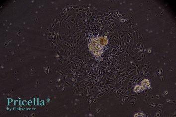
Figure 2. Cobblestone-like epithelial cell clusters
Key Considerations
1. Animal Age Matters
Kidney size and tubular structure vary with age. Choose mesh pore sizes (e.g., 100 or 200 mesh) based on animal age for better enrichment.
2. Slow Attachment Is Normal
Renal tubular epithelial cells adhere slowly—allow up to 48 hours. Collagen Type I coating significantly improves attachment and early viability.
3. Specific Nutrient Demands
Renal epithelial cells require specialized media. Standard formulations are often insufficient—use growth factor-enriched epithelial cell media to maintain morphology and function.
Prev: Is Slow Cell Growth an Abnormal Condition? How to Adjust it Properly?
Next: Decoding Astrocytes: Essential Techniques for Primary Cell Isolation


