The Black Dots in Cell Culture: What Are They?
Source: PricellaPublished: 2025-02-10
When it comes to cell culture, many researchers have encountered "black dots" in the culture medium, causing frustration and confusion due to their unknown nature. In this article, we will reveal what these black dots are and help you quickly troubleshoot your experiments.
01 Serum Precipitates
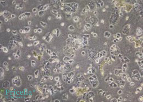
During the thawing process, serum can produce precipitates that appear as small particulate matter or even fibrous structures. These result from the precipitation of proteins and salts during serum thawing.
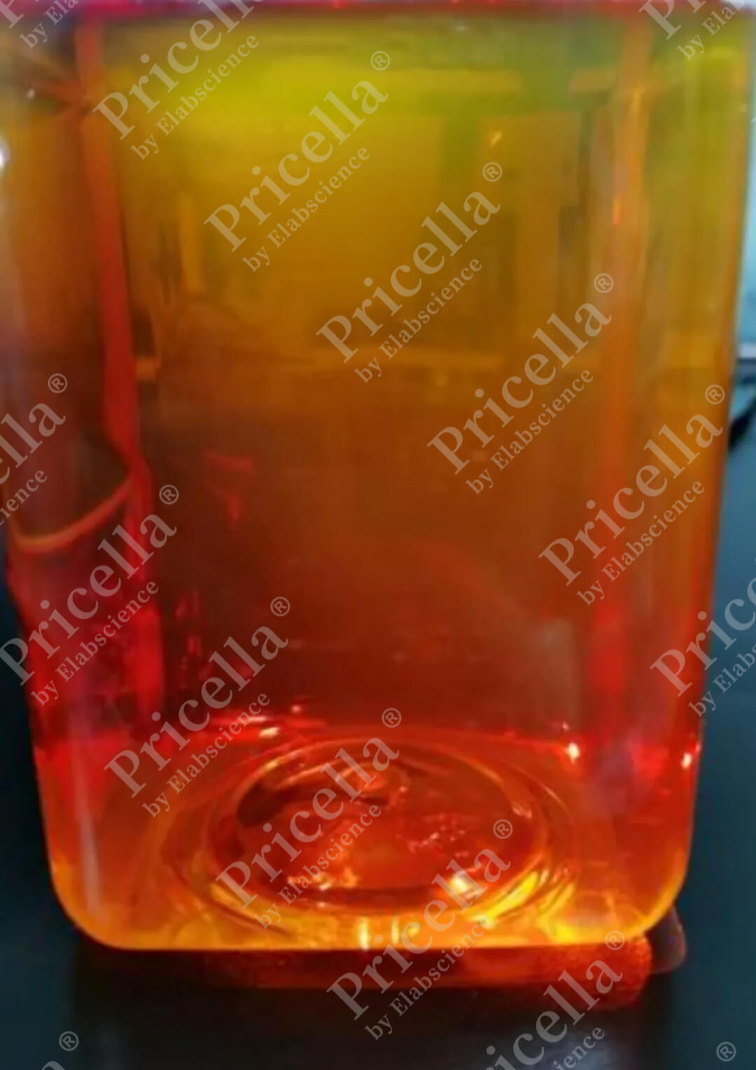
Serum was thawed with stirring and mixing without black dots.
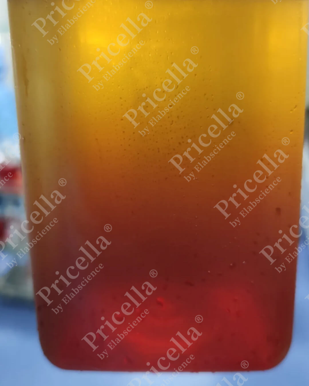
Static thawing of serum without stirring can cause noticeable black dots.
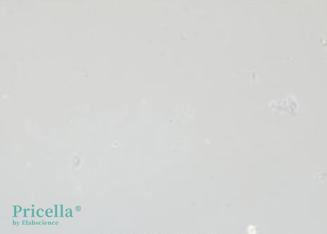
Serum Precipitates Under the Microscope
Influencing Factors
The amount of precipitate largely depends on the thawing method. Thawing serum at 4°C with continuous shaking and mixing can effectively reduce precipitate formation. In contrast, static thawing at 37°C produces more precipitates.
Solution
To address serum precipitates:
• Centrifuge the serum to remove visible precipitates.
• Failure to do so may result in an abundance of black dots and impurities during cell culture, which can interfere with observations.
02 Cell Debris or Secretions
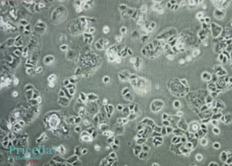
During cell metabolism, apoptotic cells release debris and secretions that appear as black dots under the microscope.
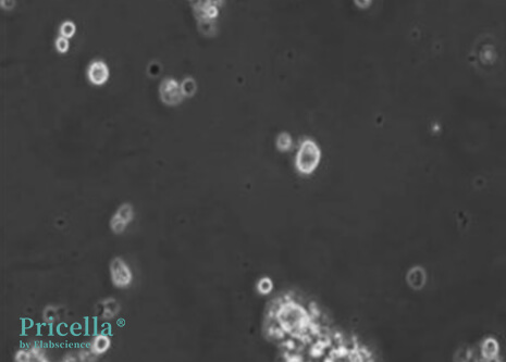
Cell Debris and Secretions Under the Microscope
Characteristics
① Size Variability: Cell debris appears as unevenly sized black dots, increasing slightly over time but not exponentially.
② Cell State Dependent: Poor cell health leads to increased debris, while improved cell conditions reduce the black dots.
③ Cell-specific Secretions: Some cell lines produce significant secretions, while others do not. Secretions typically do not impact cell morphology or proliferation rates.
03 Exogenous Microbial Contamination
Contamination with mycoplasma, nanobacteria, or bacteria can also produce many black dots. In bacterial contamination, these dots may even lead to a turbid culture medium.
In mycoplasma contamination, numerous black dots are observed.
Following the addition of the Anti-Mycoplasma Treatment Reagent, black dots significantly decreased by the next day.
How to Differentiate Between Contamination and Cell Debris?
1. Exogenous Microbial Contamination
• Mycoplasma and nanobacteria contamination are generally accompanied by a significant increase in black dots and can lead to slower cell growth, increased cell death, and poor overall cell health.
• Addition of specific removal reagents significantly improves cell condition and reduces black dots.
2. Cell Debris/Secretions
Black dots caused by cell debris or secretions persist even after adding removal reagents.
They do not influence cell growth or proliferation.


